What is Diabetic Retinopathy?

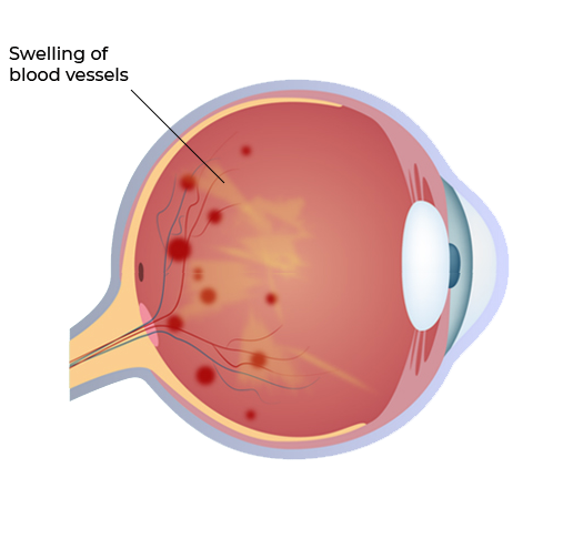
Diabetic retinopathy is a sight-threatning complication of diabetes, which is result of damage to the tiny blood vessels that nourish the retina. They leak blood and other fluids that cause swelling of retinal tissue and clouding of vision. This is "non-proliferative Diabetic Retinopathy". The condition usually affects both eyes.

If the eye disease worsens, areas of the retina may not get enough blood supply to which the eye responds by growth of new blood vessels on the surface of retina. This is termed as "proliferative Diabetic Retinopathy" characterized by complicated problems like "vitreous hemorrhage" (bleeding inside the cavity of the eye) and "tractional retinal detachment". It causes progressive damage to the retina (the light sensitive lining of the eye).

slider image

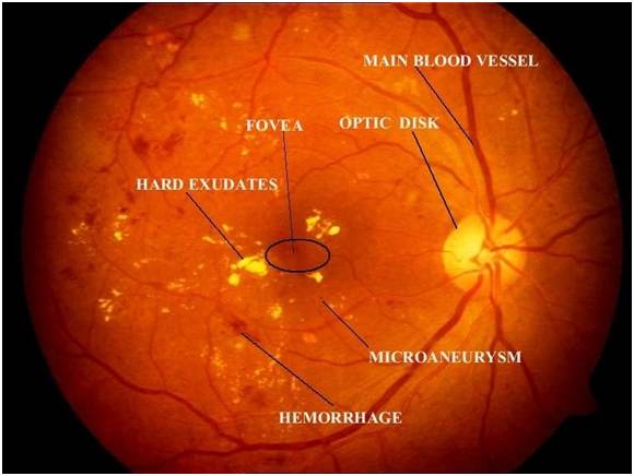
Non-proliferative Diabetic Retinopathy

slider image

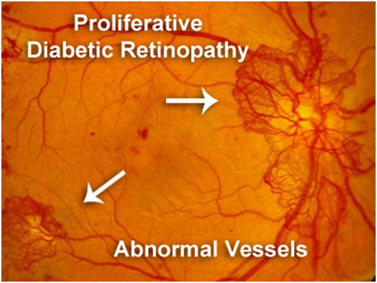
Proliferative Diabetic Retinopathy

Symptoms of Diabetic Retinopathy include


slider image
Floaters

Spots / floaters are seen in your field of vision.

slider image
Dark Spot

Vision having a dark or empty spot in the center of vision 

slider image
Blurred vision

Overall vision in unclear and blurred

slider image
Reduced Night Vision

Low light / night vision is evidently reduced

Diabetic Macular Edema

What is Diabetic Macular
Edema (DME)?


When these damaged blood vessels leak and deposit fluid over or under the macula (the central retina which is most important to carry out fine vision work), it results in swelling and termed as "Diabetic Macular Edema". 
Diabetic Macular edema is the most common cause of vision loss in people who have diabetes. 

Types of DME

There are two sub types of DME


  1. Focal DME - abnormalities in the blood vessels in the eye lead to Focal DME. It is generally observed to be milder of the two subtypes with less macular thickening, better visual acuity, and lesser degree of retinopathy severity. 

  2. Diffuse DME - It's more severe with greater degree of retinopathy severity and leads to more macular swelling and vision blur than Focal DME. 


Who is at Major Risk of DME?

Those who have had diabetes. for an extended amount of time are at high risk.


Also

  • Severe hypertension (high blood pressure).
  • Fluid retention.
  • Hypoalbuminemia (low levels Of protein in body fluids).
  • Hyperlipidemia (high levels of fats in the blood).

What investigations are needed if I have Diabetic Retinopathy?


OCT


Optical Coherence Tomography (OCT)


An OCT scan is a non-invasive latest advancement in imaging technology. Similar to ultrasound, this diagnostic technique employs light rather than sound waves to achieve higher resolution (5 micron resolution) pictures of the structural layers of the ratina very prcisely. Thus enhancing patient care. It has the ability to detect problems in the eye prior to any symptoms, providing guidance to prevent progression of the disease. 

DIA


Fundus Fluorescein Angiography (FFA)


It is a common procedure that is performed to give more information about the blood vessels of the retina. A fluorescein dye is injected in the blood vessel of the arm. Then by specialised magnifying camera quick photographs are taken of your retina. This helps retina specialist to understand and study the pathology in details, which guides the treatment process. 

Patient Reviews

VIEW ALL

Testimonials

VIEW ALL